This full-color photographic atlas is designed as a visual reference to accompany any human anatomy, human physiology, or combined human anatomy and physiology textbook or lab manual. It will be of particular value to students in the laboratory setting.
Features include the following:
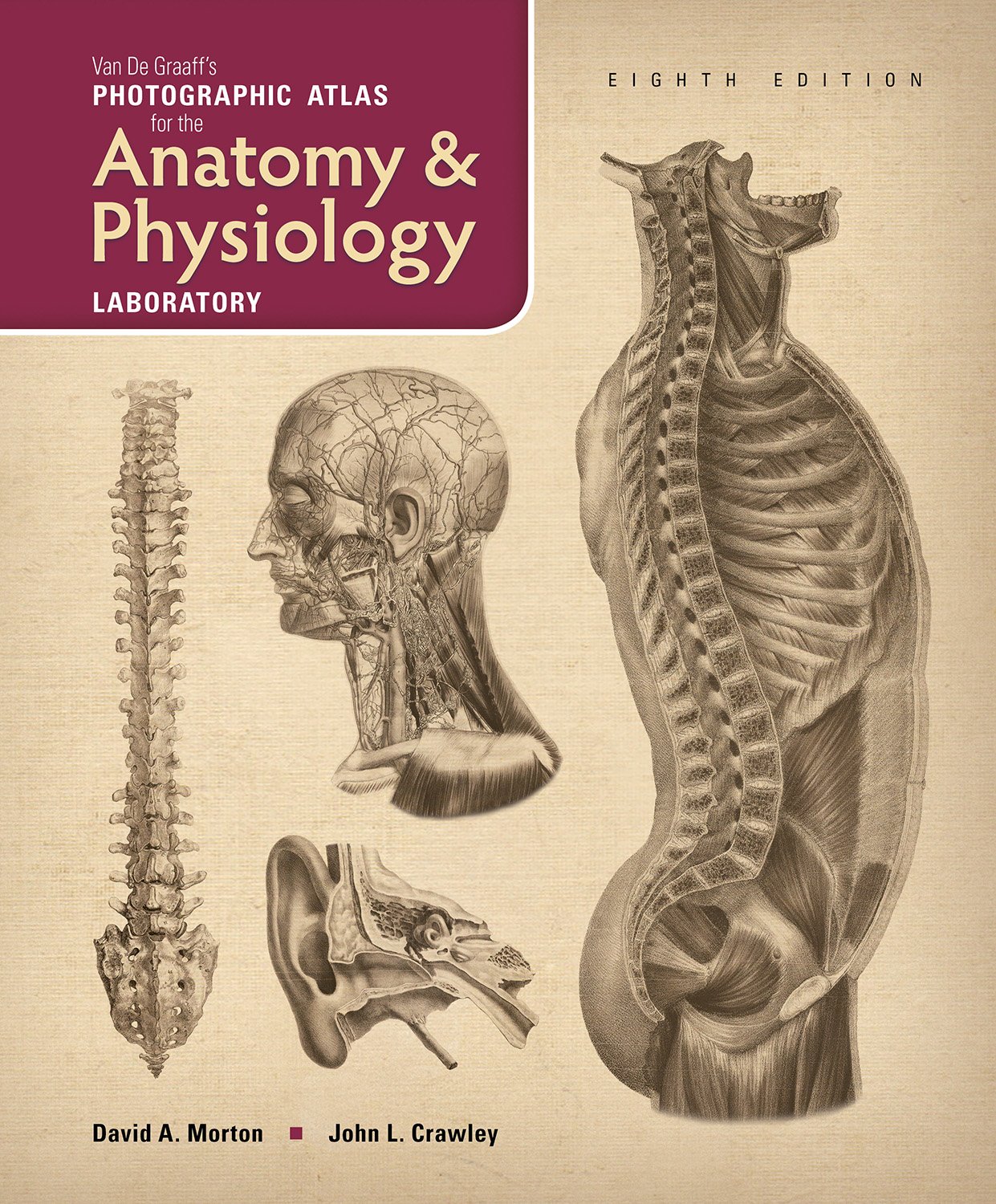
More than 600 high-quality, carefully labeled photographs, micrographs, and illustrations serve as visual references for students;
completely labeled, informative figures are depicted clearly and accurately;
multiple images of the muscular, skeletal, and other organ systems provide a complete picture of the layers of human anatomy;
microanatomy is presented as a guide for students microscope work in the lab;
photographs of sheep heart, brain, and eye dissections are included;
cat, fetal pig, and rat dissections are included
Van De Graaff's Photographic Atlas for the Anatomy & Physiology Laboratory, 8e
📄 Viewing lite version
Full site ›
29.91
USD
🛒 Buy New on Amazon 🇺🇸
Book Details
Author(s)David A. Morton, John L. Crawley,
PublisherMorton Publishing Company
ISBN / ASIN1617312770
ISBN-139781617312779
Sales Rank19,158
MarketplaceUnited States 🇺🇸
Description ▲
Similar Products ▼
- Fearfully and Wonderfully Made
- Exploring Anatomy & Physiology in the Laboratory, 3e
- An Illustrated Atlas of the Skeletal Muscles
- Hole's Human Anatomy & Physiology, 13th Edition
- A Visual Analogy Guide to Human Anatomy & Physiology, 3e
- Exercises for the Anatomy & Physiology Laboratory, 3e
- Anatomy & Physiology: The Unity of Form and Function
- The Anatomy Coloring Book
- Human Anatomy & Physiology (Marieb, Human Anatomy & Physiology) Standalone Book
- Principles of Human Anatomy